Super LSI
Superior Large-area
CMOS Technology
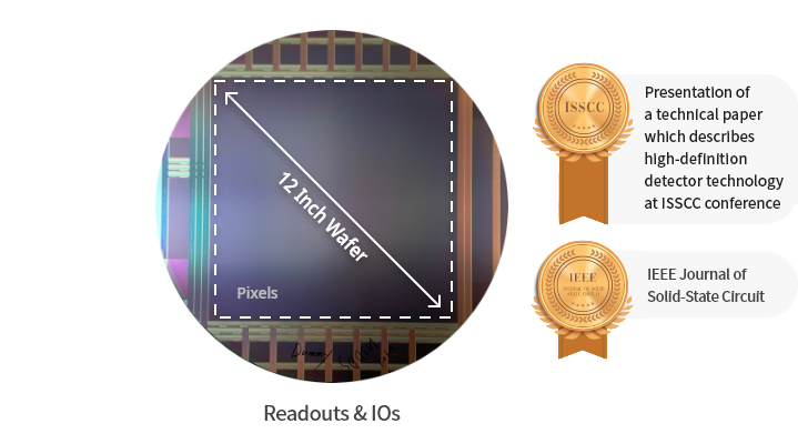
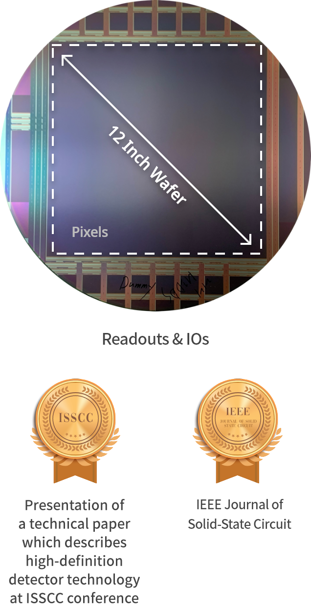
Super LSI realized the largest 12-inch CMOS in the world with Rayence’s large-area wafer technology, unlike existing small and medium sized CMOS sensors. You can acquire a high-resolution premium image with a fine pitch design. And, you can reduce noise to create X-ray images of a different level by applying on-chip ADC design.
Video of MIDAS 2121 to which Super LSI CMOS technology is applied
ADC on Chip
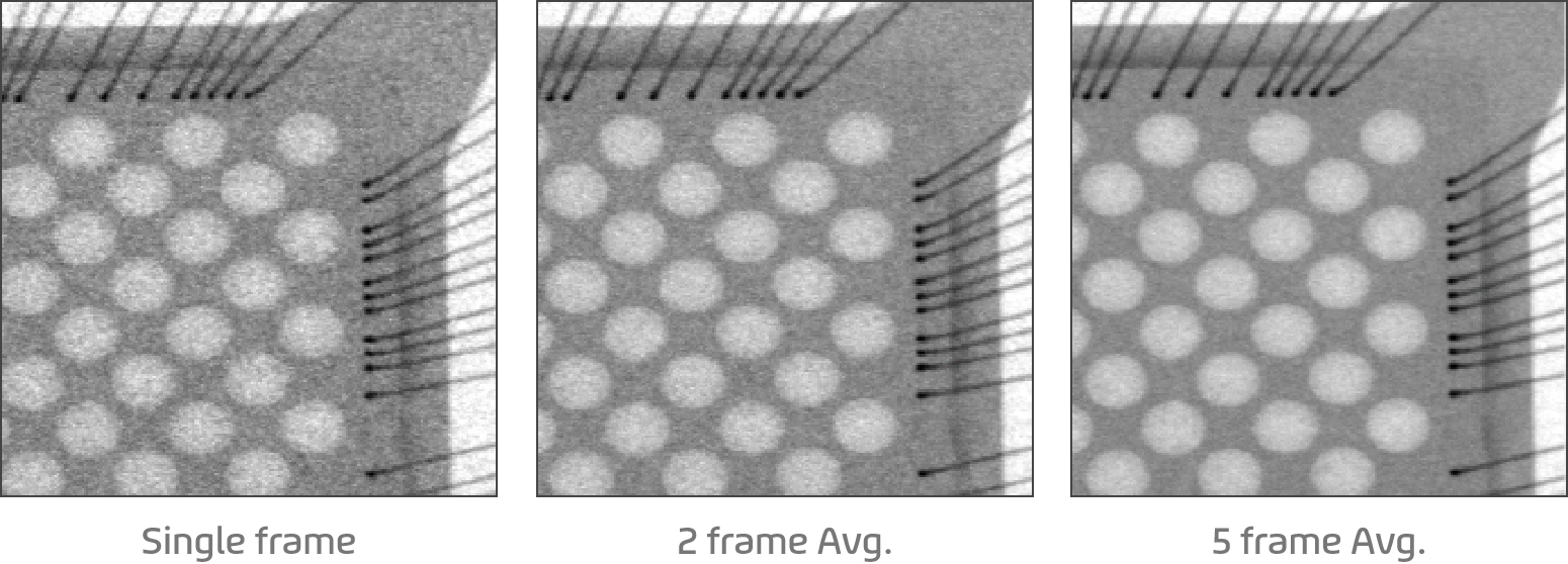
Applies ADC on chip design that converts analog signals to digital signals inside CMOS and outputs them to the outside. Outputs the digitally converted value to the outside of the chip to minimize the noise inflow path to the signal, thereby creating low noise (32µVrms). Carries out digital conversion of analog signals in parallel in 4,608 columns to implement high speed readout (14µs/row).
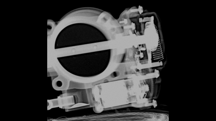
rayence MIDAS 2121
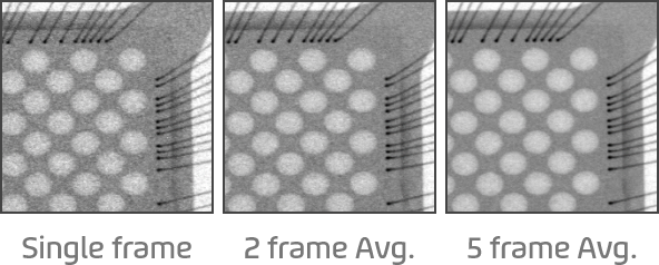
Competitor
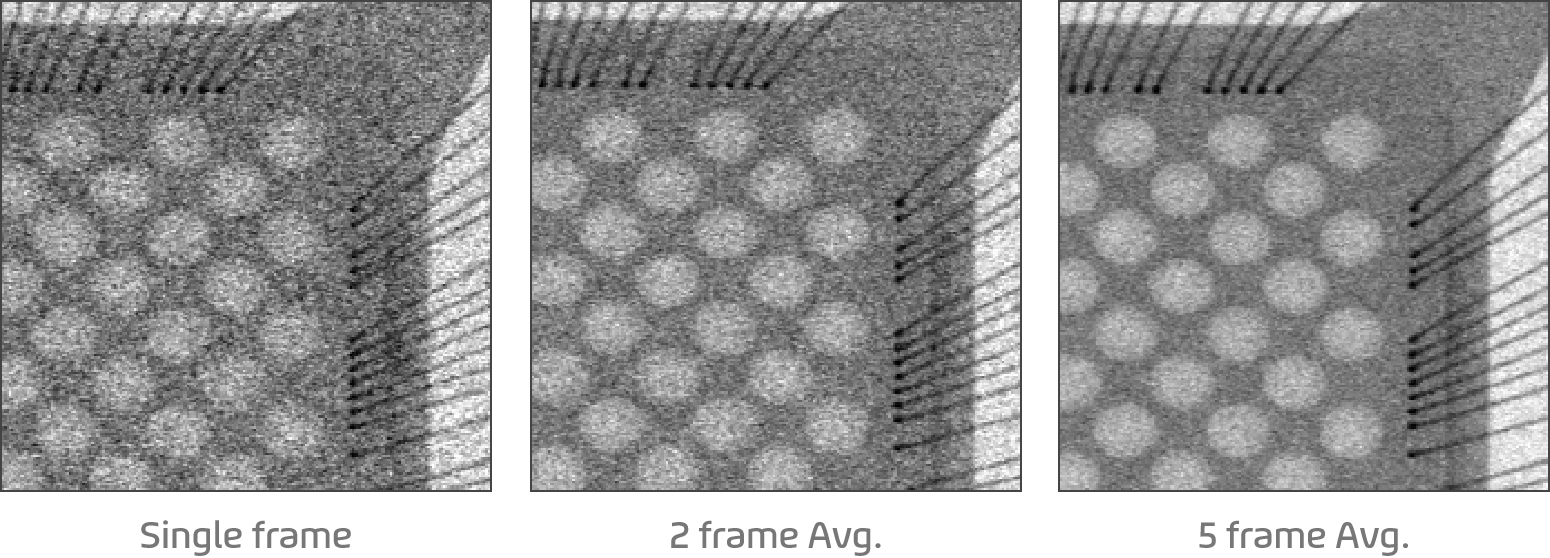
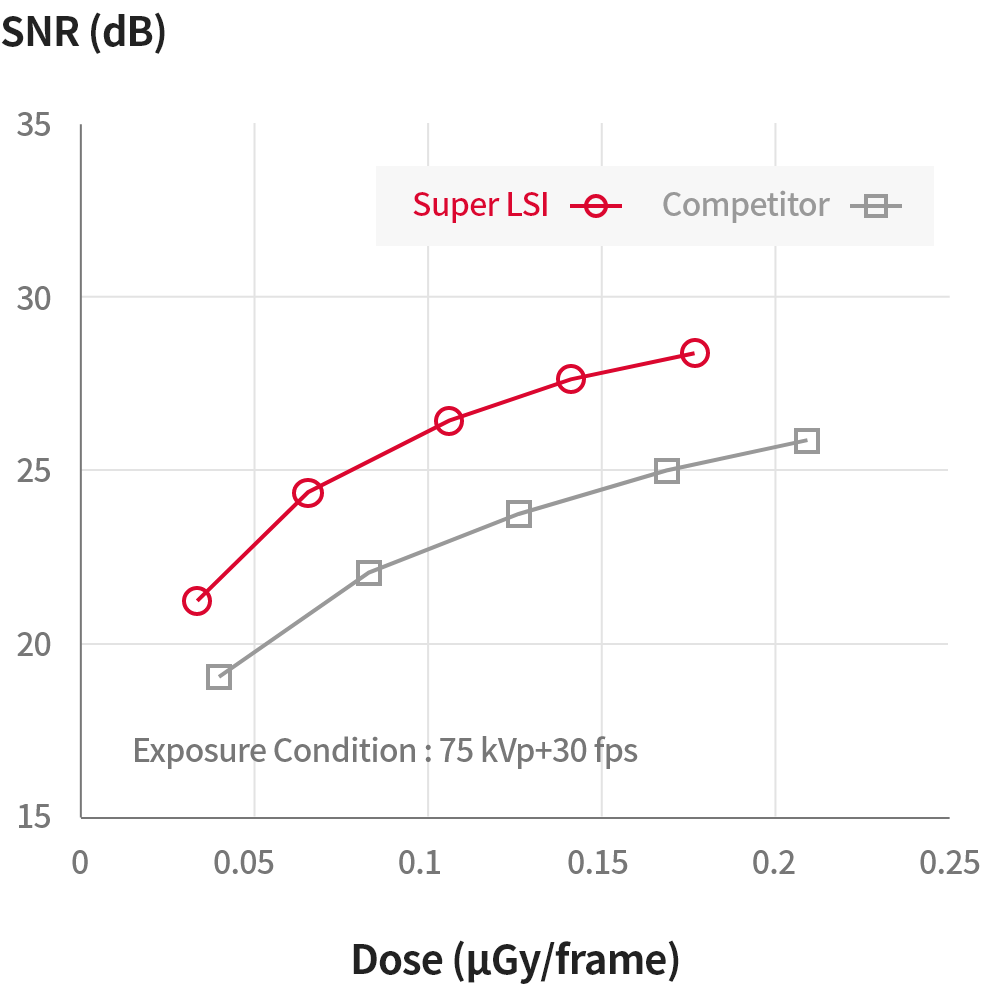
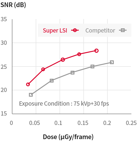
Achieves excellent SNR characteristics improved by about 12% compared to competitors by reducing noise through ADC on chip.
* The chip converts the analog signals collected from the pixel to digital from inside and then outputs to the outside to minimize the noise inflow path.
Super IGZO TFT
Enhanced the image acquisition speed compared to a-Si TFT
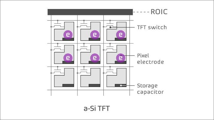
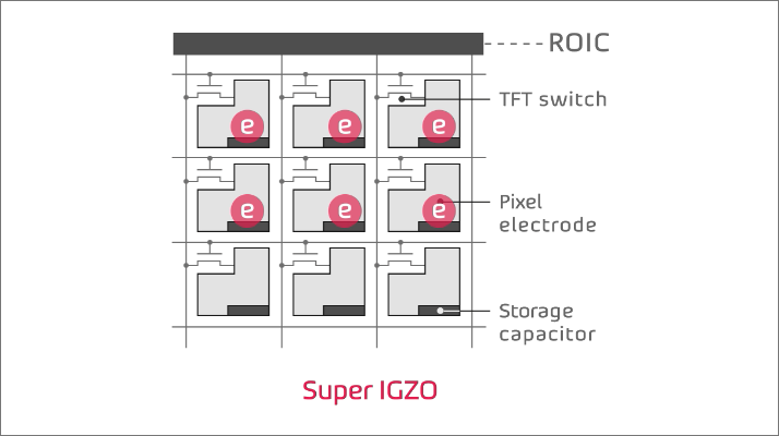
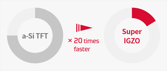
Image lag occur as it will remain in the pixel.
Carrier mobility of Super IGZO is 20 times
higher than that of an a-Si TFT
In addition, Super IGZO is a Rayence’s self-development technology that improves image acquisition speed by more than 20 times compared to a-Si TFT. Especially, Super IGZO improves image quality and work efficiency in the applications that require fast image acquisition, such as X-ray imaging for human body surgery, panorama/CBCT imaging for dental, and industrial in-line inspection.
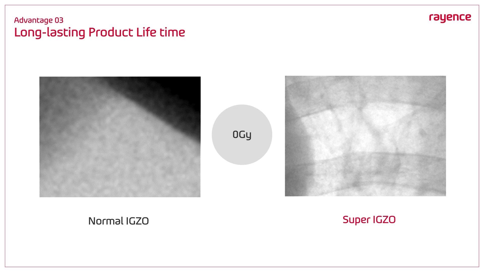
World First Hardness up to 10,000 ㏉
In addition, Super IGZO is a Rayence’s self-development technology that improves image acquisition speed by more than 20 times compared to a-Si TFT. Especially, Super IGZO improves image quality and work efficiency in the applications that require fast image acquisition, such as X-ray imaging for human body surgery, panorama/CBCT imaging for dental, and industrial in-line inspection.
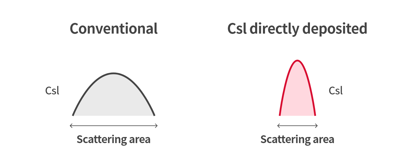
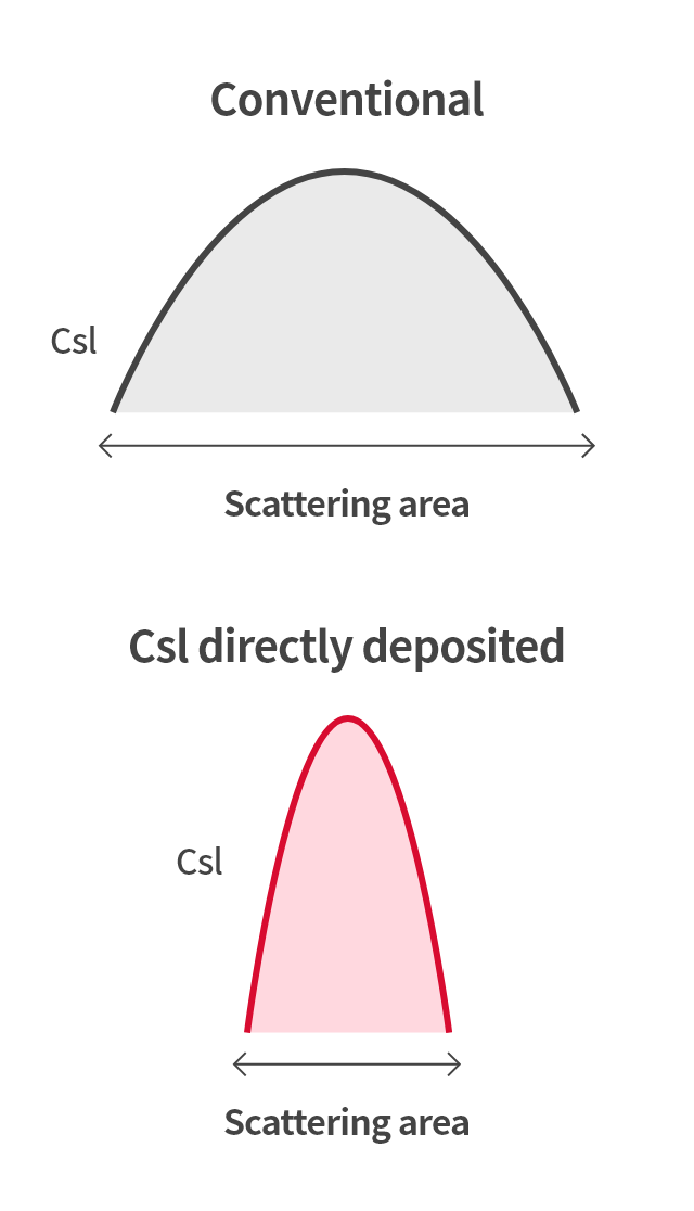
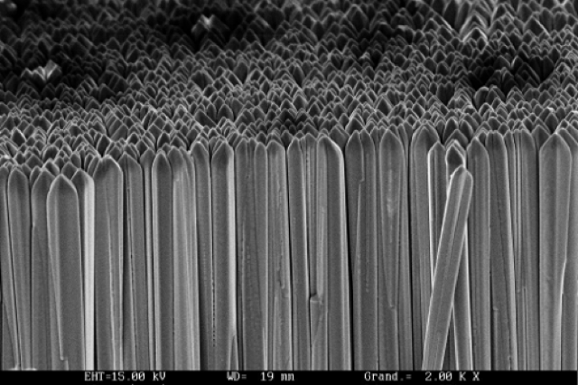
Specialized Csl Technology
Enhanced Image Quality
The core technology of Rayence CsI is to minimize X-ray scattering. In the process of converting X-ray into visible light to be moved through the scintillator to the pixel, the direction of X-rays is deviated. That is, scattering of X-rays occurs. As a solution for this, we developed a Low Dose CsI technology which minimizes scattering. We improved the blurring phenomenon to increase the contrast and secure high resolution while minimizing an area where scattering occurs.
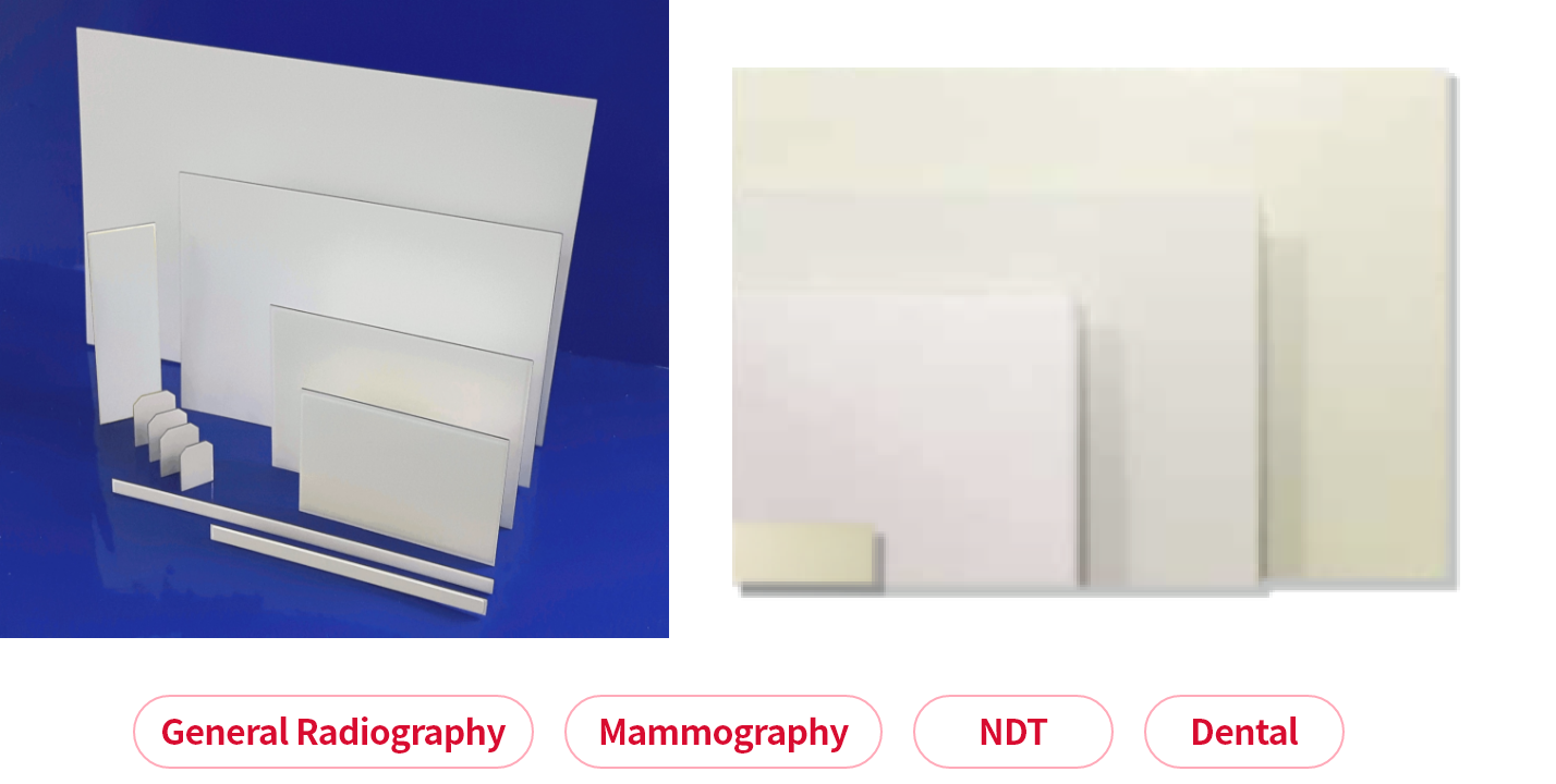
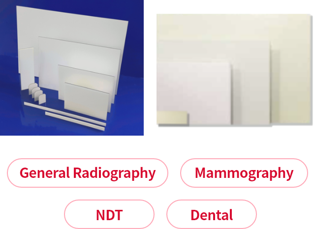
Bespoke Application & Dimension
We develop and supply a scintillator which meets the X-ray applications and customer's requirements. We provide the optimal solutions and services so that you can concentrate on various X-ray inspection businesses that customers require, such as medical, breast examination, dental, and non-destructive inspection.
Excellent Uniformity
With in-house deposition technique considering the characteristics of CsI, in which the material structure is arranged alone, we provide the excellent quality image even at a low dose. We can convert X-rays into electrical signals to create images stably and provide the best image quality in its class.
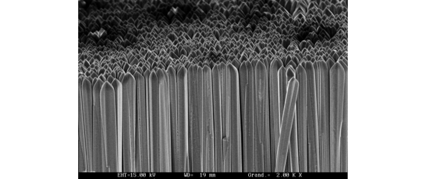
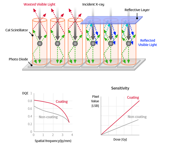
Superior to-the-edge coating
Since we apply a reflective layer to improve sensitivity, it is possible to develop a product according to a customer’s requirements, such as moisture prevention and creation of various thicknesses of the scintillator. We have upgraded CsI technology further by targeting video performance indicators such as Sensitivity, DQE, and MTF that affect video quality.

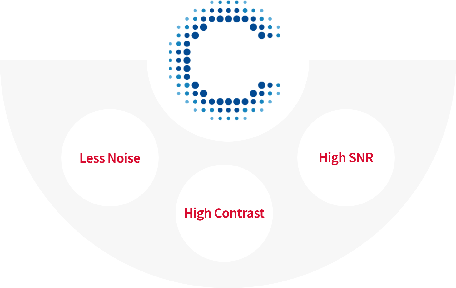
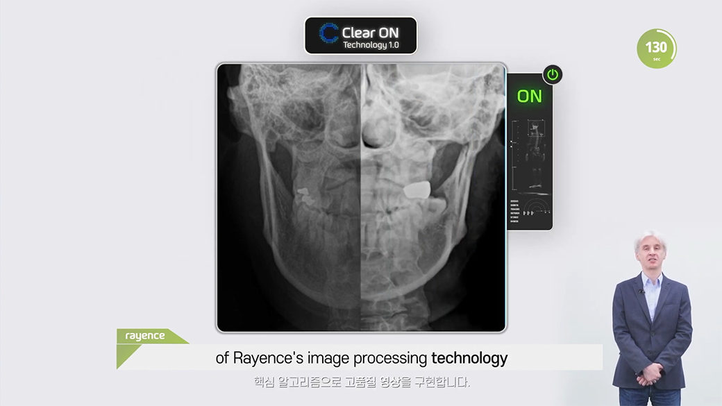
Clear ON
Enhanced Image Processing for Diagnosis Accuracy Improvement
Clear ON, the main algorithm of Rayence image processing provides clear X-ray images. Clear ON expands the range of the gray scale to increase the contrast and sharpness of the image. In particular, Clear ON is an intelligent algorithm specialized for medical X-ray imaging, and you can pay attention to the performance improvement of HDR and super resolution.
It was difficult to read the soft tissue area before HDR image processing, but you can see the image clearly without a missing part in the X-ray images processed with HDR. Therefore, you can display various areas at once without omission of soft tissue, thereby enabling image processing optimized for reading.
The advantage of improving performance of super resolution is that you can increase the resolution of an image to create high resolution image. Because it is possible to read clinical images with detailed data, you can detect lesions or defects quickly and accurately.
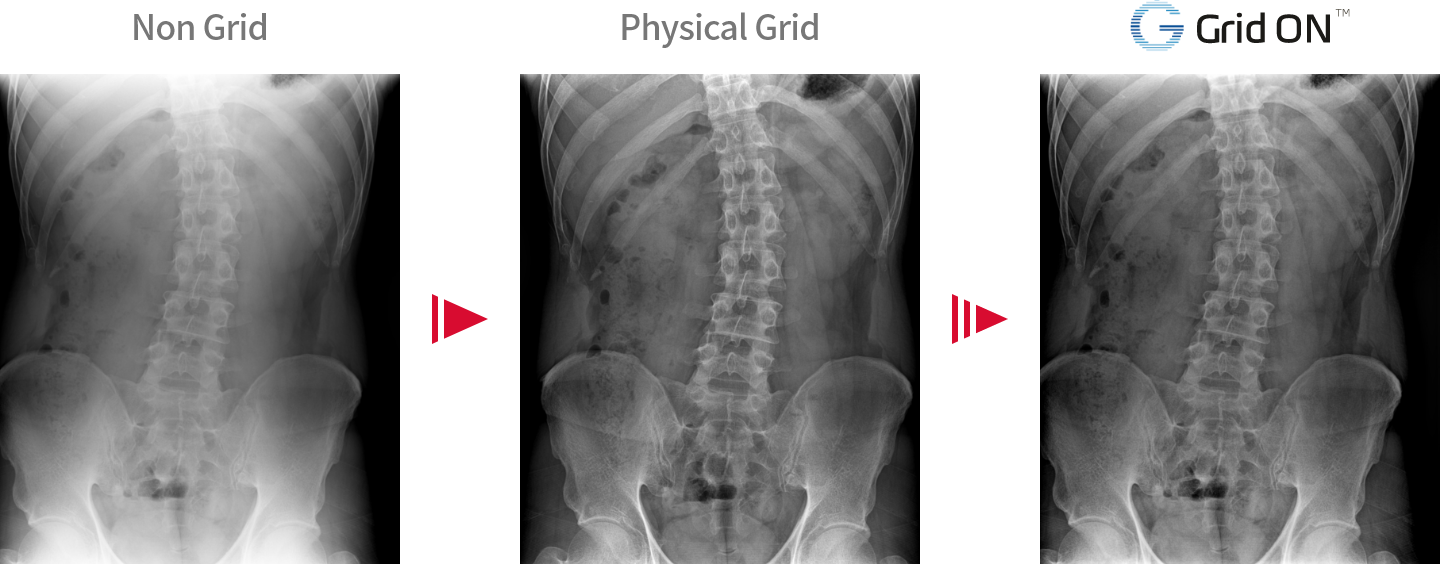
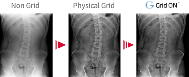
Grid ON
Anti-Scatter Algorithm
Grid ON is a Virtual Grid algorithm to reduce scattered X-rays.
This is an Anti-Scatter technology that implements the grid function in SW without a grid, and you can shoot with a lower dose.

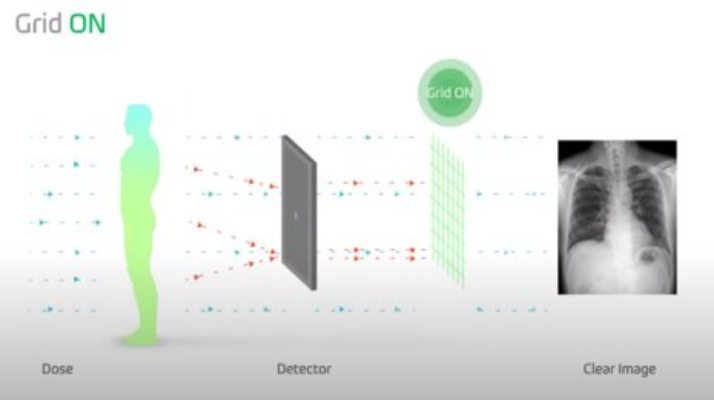
The Grid ON, to which the anti-scatter algorithm is applied, plays the role of virtual grid that removes scattered X-rays and provides an acquired X-ray image as a high-quality image.
Images, to which Grid ON is applied, have improved sharpness and contrast to support more accurate reading.
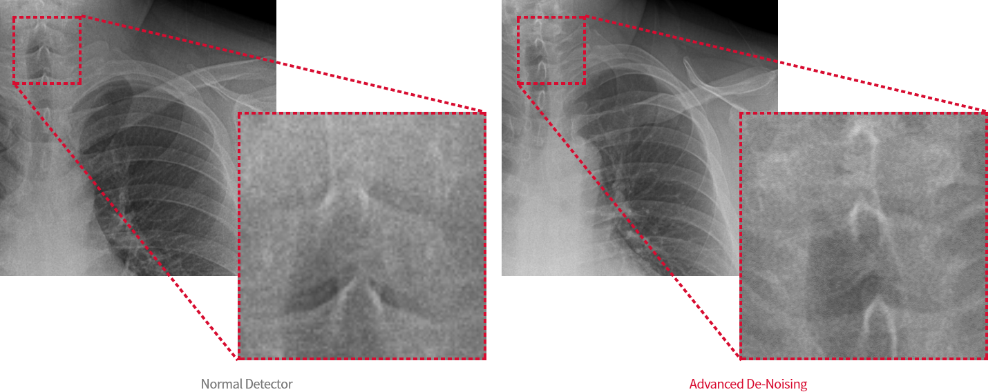
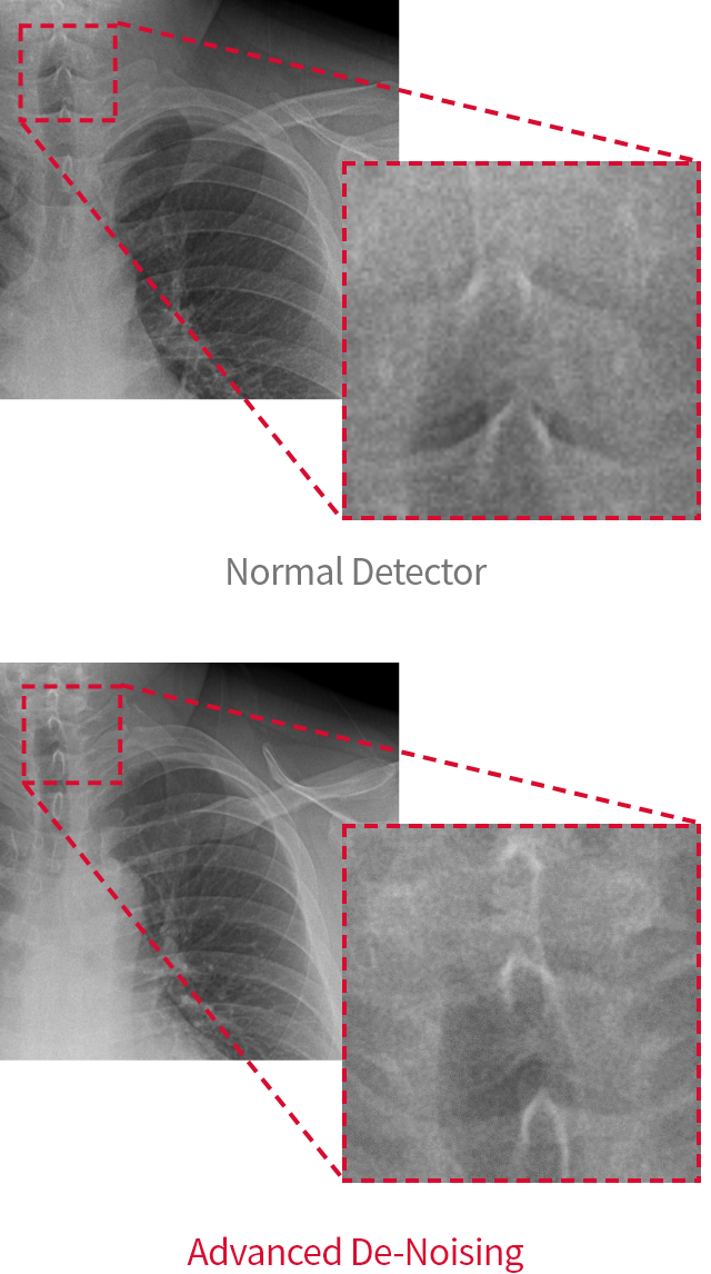
Advanced De-Noising
Noise Reduction Image Processing
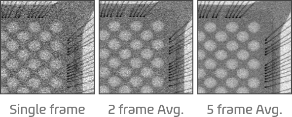
Advanced De-Noising is a solution developed to reduce noise, which is of the main causes of hindrance to reading. Advanced De-Noising minimizes the noise generated when converting X-rays into electrical signals and improve the existing De-Noising algorithm to provide excellent SNR* performance even at low doses.
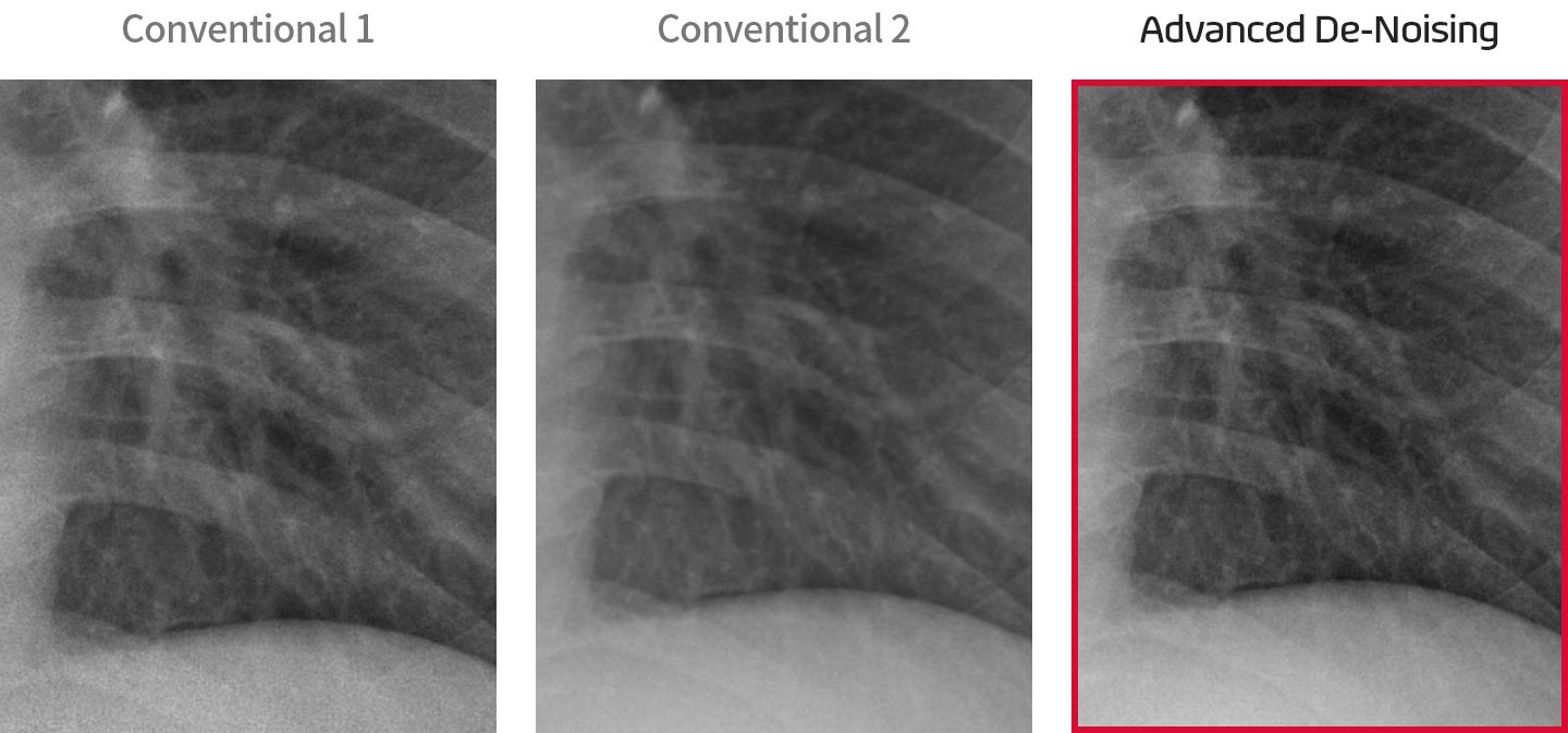
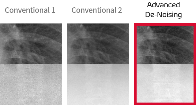
This increases the sharpness with the Advanced De-Noising algorithm to detect microlesions and abnormal signs very well, and continues to improve the noise reduction algorithm, thereby realizing image quality and high performance. Since this identifies the noise in the acquired image, image processing is possible to improve the accuracy of reading.
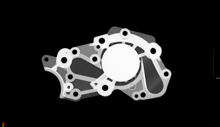
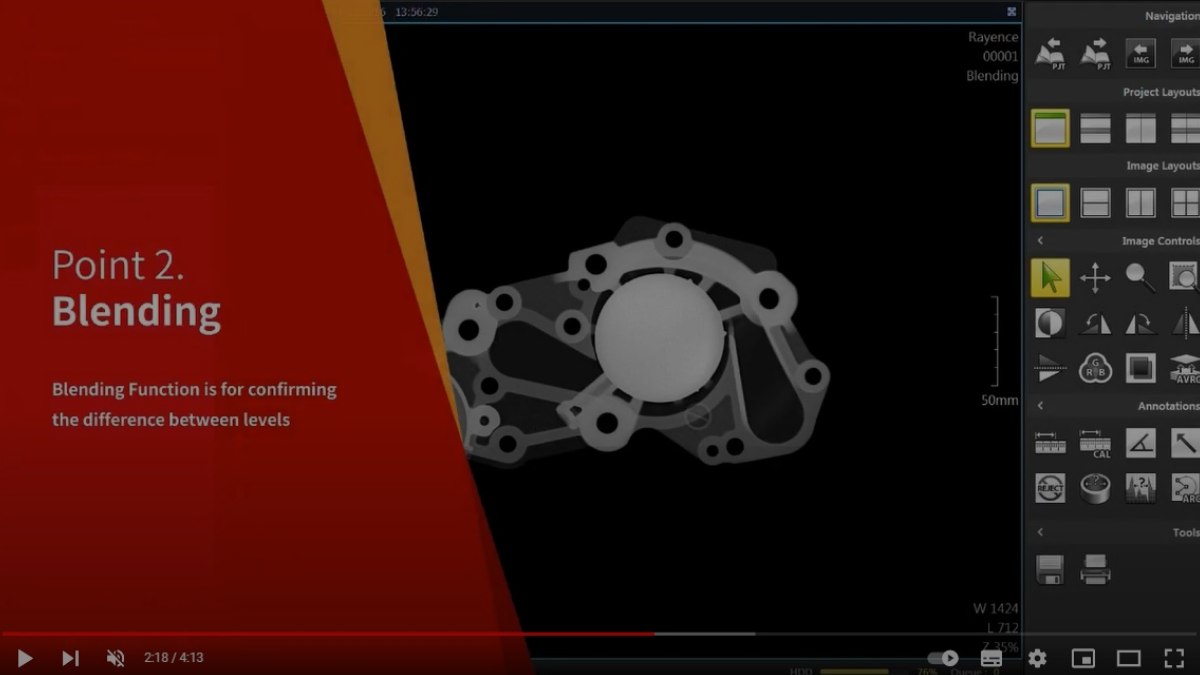
Blending
Image Processing for Object of Different Level
Blending is an image processing algorithm that is useful for portable NDT X-ray inspection and is a rayance proprietary technology.
Blending has been developed to inspect subjects with different level values. You can acquire shootings of different level values from one subject, then combine them in the same position to read them at once with this technology. And, this increases work efficiency in industries where inspection time is directly related to productivity.
Medical Screening Solution
Imaging Recognition
Technology
This detects the findings of major lesion automatically when reading chest X-ray images to improve the efficiency and accuracy of diagnosis.
In addition, it provides a support for a medical staff to make quick and accurate decisions in the X-ray imaging environment.
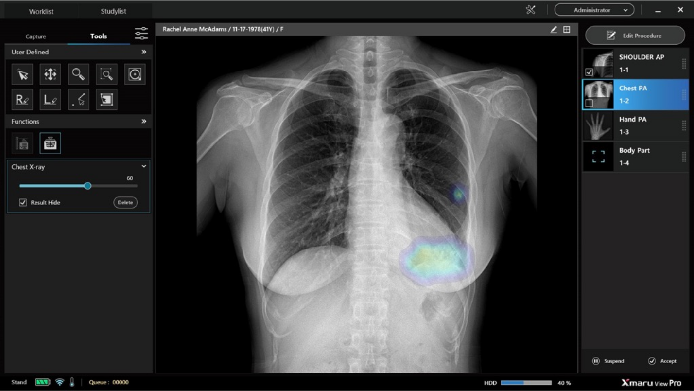
Assist with image reading through learning of major abnormal findings identified in chest X-ray images
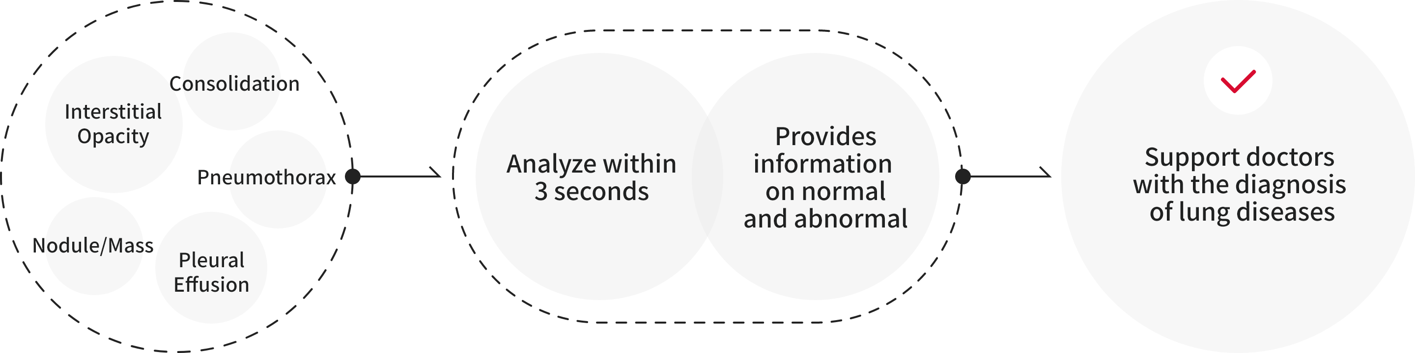
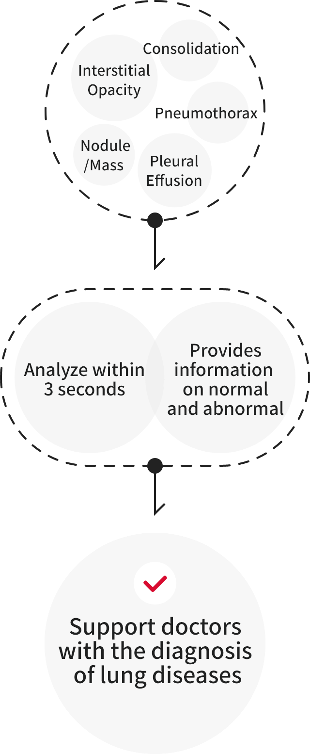
It provides normal, abnormalities analysis and information for the five major findings, and a combination of the findings provided to assist in the treatment of major lung diseases such as lung cancer, tuberculosis, and pneumonia.
Imaging Recognition
Technology
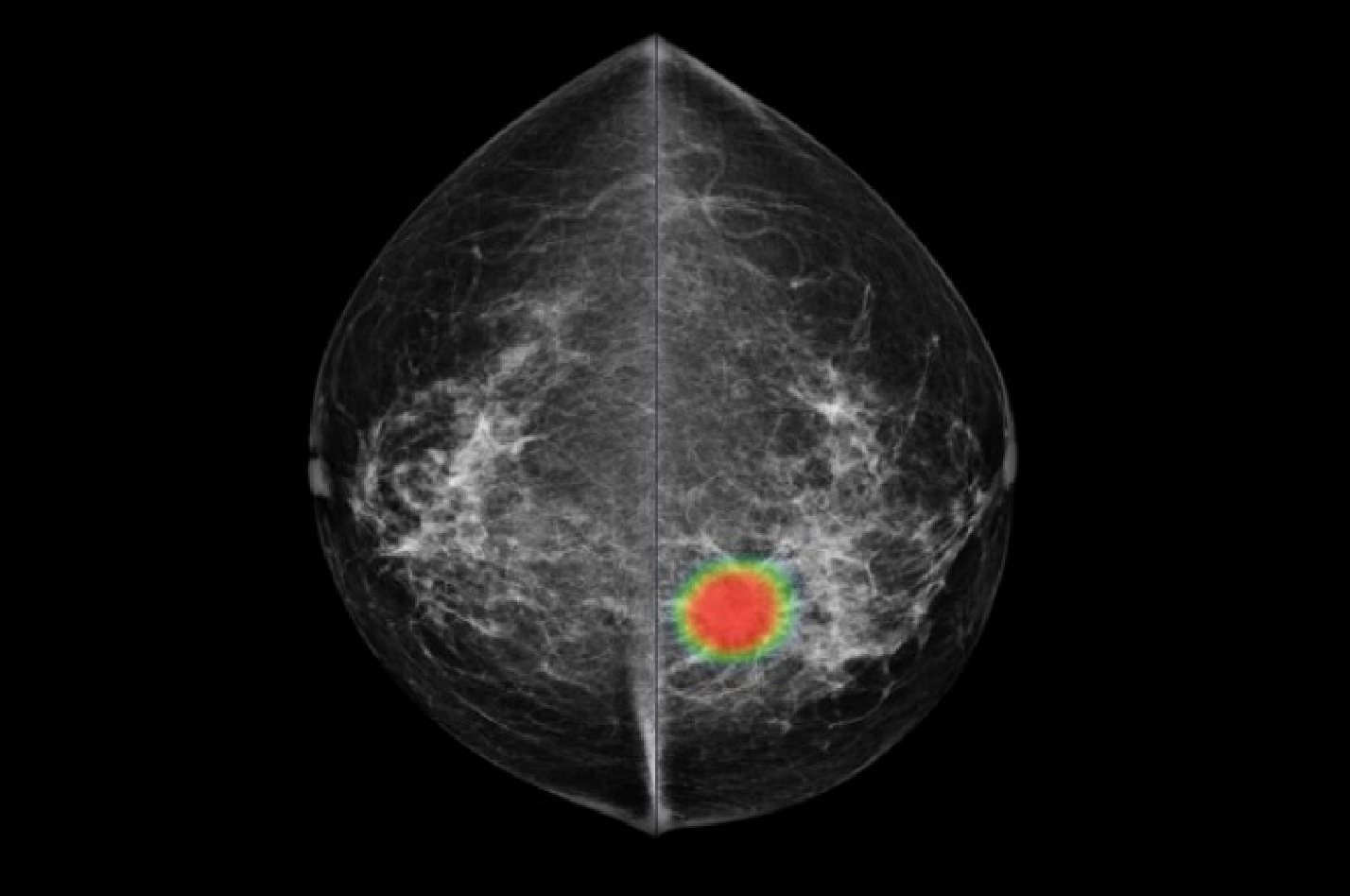
This is assisted diagnosis solution specialized for breast cancer detection. The solution improves the accuracy of breast cancer diagnosis, thereby increasing the likelihood of early detection of breast cancer. In addition, the solution improves the mammogram performance significantly.
Mammo Insight Delivery Information
Displays the areas suspected of breast cancer with heat maps and outlines.
Derives the probability of the breast cancer for each breast as a probability value.
Classifies breast density into 4 categories.
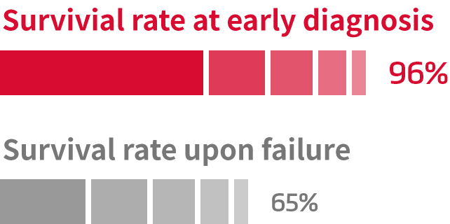
Improves survival rate at early diagnosis of breast cancer.
Five-year survival rate (Survival rate at early diagnosis vs Survival rate in case of failure)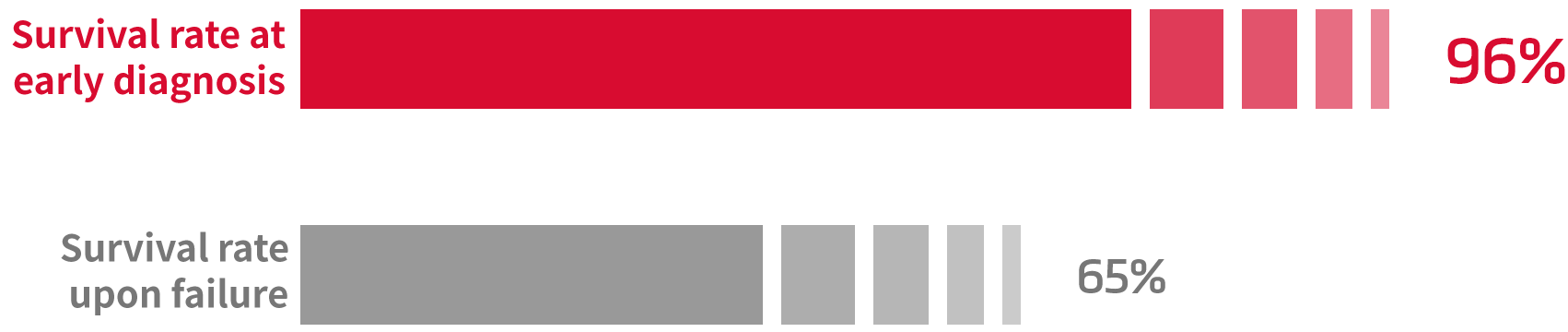
Imaging Recognition
Technology
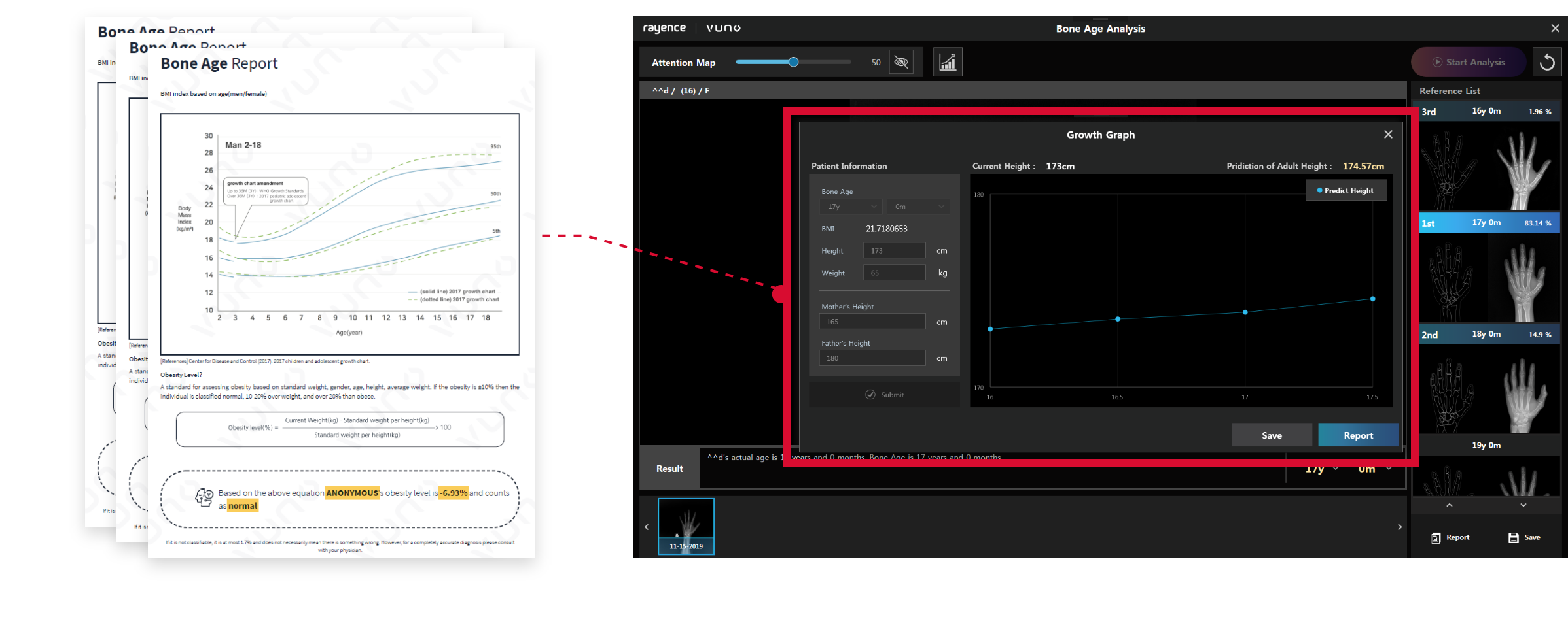
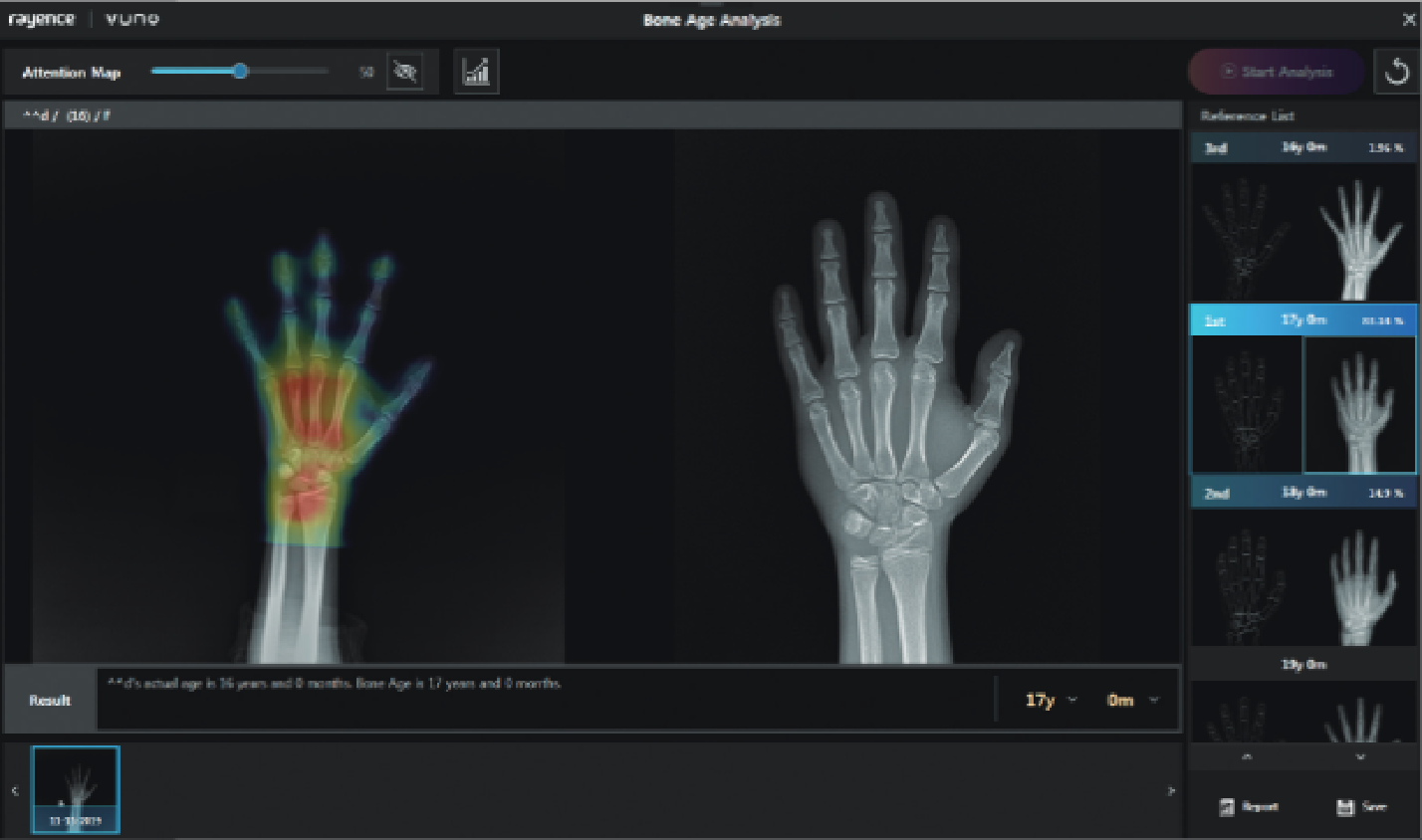
This technology analyzes X-ray images to provide up to the top 3 rankings of the most similar bone ages among the age groups (31 males, 27 females) defined by the GP method (Greulich-Pyle). And, this technology is an auxiliary solution that provides analysis results within a few seconds with a single click for supporting medical staff’s reading.
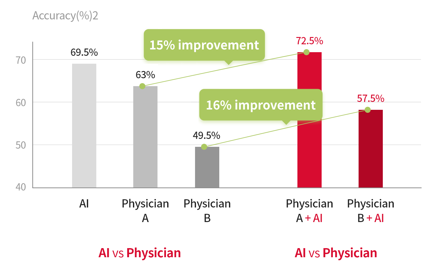
The concordance rates increased with the use of this Imaging Recognition Technology.
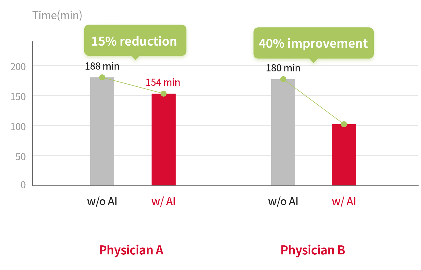
Imaging Recognition Technology allowed us to reduce the total reading times of the radiologists.
References 1. Computerized Bone Age Estimation Using Deep Learning-Based Program: Evaluation of the Accuracy and Efficiency AJR:209,December 2017
References 2. Total reading time of bone age based on 200 X-ray images of left-hand bones
Providing Bone Age report
through the growth chart